
Managing Hematocrit and Erythrocytosis on TRT: A Practical Monitoring Guide (2026)

If you are on testosterone replacement therapy, hematocrit is the lab value most likely to interrupt your protocol. It is the single most common reason a clinician pauses a dose, switches a route, or sends you for a blood draw, and yet it usually causes no symptoms at all, which is exactly what makes it a monitored-lab problem rather than a felt one. The good news is that high hematocrit on TRT is predictable, trackable, and in almost every case manageable without abandoning therapy. This guide explains what hematocrit is, why testosterone pushes it up, the thresholds clinicians actually act on, and the evidence-ranked toolkit for bringing it back down, including the 2024 research that complicates the old "just donate blood" reflex.
A quick honesty note: this is a "what is happening and what gets done about it" guide, not a hematology textbook and not a how-to. Every threshold and dose figure here is a guideline number or a typical clinic range, presented for context, and every intervention, from a dose change to a blood draw, is a clinician's decision. Where a topic runs deep, like injection-frequency mechanics or the full bloodwork panel, we summarize and link to its dedicated guide so this page stays focused on the one job it owns: hematocrit.
Key Takeaways
- Hematocrit is the fraction of your blood made of red cells, and a normal range runs to roughly the high 40s in percent; testosterone reliably raises it, a side effect called secondary erythrocytosis (not the bone-marrow cancer polycythemia vera).
- The thresholds that trigger action are layered. Many clinicians start watching closely near 52%, the Endocrine Society defines erythrocytosis at >54% and advises pausing testosterone until it normalizes, then resuming at a lower dose. A 2024 review notes the 52/54% cutoffs are weakly evidenced.
- Dose reduction and smoother levels come first. Lowering the peak (smaller, more frequent doses, or switching from intramuscular to subcutaneous or gel) is the first-line lever per the 2024 evidence, ahead of routine phlebotomy.
- Therapeutic phlebotomy works but is a double-edged sword. It drops hematocrit fast, but repeated draws deplete iron and may, paradoxically, keep red-cell production switched on; it is a clinician-decided procedure, not a DIY fix.
- It is usually silent, so it is a lab thing. In our tracking data the median hematocrit sits near 48% (in range), but about 9% of trackers cross the ~54% line that prompts action, which is why scheduled CBCs matter.
What is high hematocrit (erythrocytosis) on TRT?
High hematocrit on TRT means too large a fraction of your blood is red cells, a condition called secondary erythrocytosis that is a known, expected response to testosterone, and it is a different thing from the blood cancer polycythemia vera. Hematocrit is the percentage of blood volume taken up by red cells. It moves with two related numbers on your complete blood count (CBC): hemoglobin (the oxygen-carrying protein) and red blood cell count. When testosterone drives more red-cell production, all three tend to rise together.
The word people reach for is "polycythemia," but the precise term matters here. Polycythemia vera is a clonal bone-marrow disorder driven in most cases by a JAK2 mutation, and it is uncommon and serious. What testosterone causes is secondary erythrocytosis, meaning the marrow is responding normally to an outside stimulus (the testosterone) rather than misbehaving on its own. There is also a third category worth knowing: spurious or relative erythrocytosis, where the red-cell mass is normal but the plasma volume is low, usually from dehydration, so the hematocrit reads high simply because the blood is concentrated. A morning lab drawn after coffee and no water can nudge the number up on its own, which is why a single high reading is usually repeated, ideally hydrated, before anyone acts on it.

Distinguishing these matters because the response differs. Secondary erythrocytosis from TRT is managed by adjusting the testosterone and, when needed, removing blood. Spurious erythrocytosis is fixed by rehydrating and rechecking. True polycythemia vera needs a hematologist. The general candidacy and diagnosis story for TRT itself lives in our complete beginner's guide to TRT, so this page assumes you are already diagnosed and on therapy, and focuses on the red-cell side.
Why does TRT raise hematocrit?
Testosterone raises hematocrit through several overlapping mechanisms: it stimulates the kidney's erythropoietin signal, it suppresses hepcidin so more iron is freed for red-cell production, and it directly nudges the marrow, and the size of that effect tracks with how high your testosterone peaks. This is the most consequential section for actually managing the number, because the mechanism is what every intervention targets.
The starting point is erythropoietin (EPO), the hormone the kidneys release to tell the bone marrow to make red cells. Testosterone resets the relationship between EPO and hematocrit, so the body behaves as if it wants a higher red-cell mass than it did before therapy. At the same time, testosterone suppresses hepcidin, the master regulator of iron. Lower hepcidin means more iron is absorbed and released from storage into circulation, and since iron is the rate-limiting raw material for hemoglobin, freeing it up lets the marrow build red cells faster. Layered on top, testosterone appears to act on the marrow's red-cell precursors fairly directly. The net result is a reliable, dose-related rise in hematocrit that typically becomes measurable within the first few months and tends to plateau later in the first year.
The detail that turns mechanism into a management strategy is peak dependence. The red-cell response is driven not just by your average testosterone level but by how high it spikes. A long-acting intramuscular ester injected in a single large weekly dose produces a tall peak in the days after the shot, and that peak is a stronger erythropoietic stimulus than the steadier curve you get from smaller, more frequent doses or from transdermal gels. This is why route and frequency, not just total dose, sit at the center of management. The men most likely to run into trouble are therefore those on higher doses, on infrequent large intramuscular injections, and those with pre-existing risk factors such as untreated obstructive sleep apnea, smoking, chronic lung disease, or living at high altitude, all of which independently raise red-cell production.
It is worth dwelling on the iron link, because it is the piece most explanations skip and the piece that explains why phlebotomy is more complicated than it looks. Hepcidin is the body's iron gatekeeper: when it is high, iron is locked away in storage and absorption from food slows; when it is low, iron flows freely into the bloodstream and toward the marrow. By suppressing hepcidin, testosterone effectively opens the iron gates, and abundant available iron is precisely what the marrow needs to keep building hemoglobin-rich red cells. That is also why testosterone-induced erythrocytosis behaves differently from a simple "more EPO" model: the limiting factor for red-cell production is often iron, and testosterone removes that limit. It explains a clinical paradox that frustrates patients, why draining blood (and therefore iron) can sometimes fail to durably control hematocrit, a point we return to in the phlebotomy section.
One more nuance separates testosterone-induced erythrocytosis from a worrying primary disorder. In secondary erythrocytosis driven by an outside stimulus, the rise is proportionate to that stimulus and reverses when the stimulus is reduced, which is exactly why dose and route are such effective levers. The marrow is doing its job, just turned up too high. That distinguishes it from polycythemia vera, where the marrow over-produces on its own regardless of the EPO signal, and it is the conceptual reason the first move in management is to turn down the testosterone signal rather than to attack the red cells directly. The deep mechanics of injection frequency and split dosing are covered in our TRT dosing protocols and injection frequency guide, and the ester and route comparison in our testosterone esters compared guide; here the point is simply that flatter levels mean a smaller red-cell stimulus.
What hematocrit is too high? The thresholds that trigger action
There is no single magic number, but the working thresholds are layered: a baseline hematocrit above roughly 48 to 50% is a caution before starting, many clinicians intervene early around 52%, and the Endocrine Society defines erythrocytosis at greater than 54%, the level at which it advises pausing testosterone until the value normalizes. Understanding why there are several lines, and how soft they are, is more useful than memorizing one cutoff.
The guideline anchor is clear. In its 2018 clinical practice guideline, the Endocrine Society advised against starting testosterone in a man whose baseline hematocrit is already high (above roughly 48 to 50%), and recommended that if erythrocytosis develops on therapy, the clinician should withhold testosterone until hematocrit returns to normal, then restart at a lower dose. It set the working definition of erythrocytosis at a hematocrit above 54% (Endocrine Society, 2018, "Testosterone Therapy in Men With Hypogonadism", retrieved 2026-06-17). That 54% line is the one most TRT clinics treat as the "stop and reassess" threshold.
In practice, many clinicians act earlier, often around 52%, on the logic that it is easier to nudge a trend down than to reverse a value that has already crossed the line. This earlier-intervention habit is widespread, but it is worth being honest that it is a clinical convention rather than a validated cutoff. A 2024 review pointed out that the 52% and 54% thresholds are only weakly supported by outcome evidence, that the link between a modestly elevated hematocrit and actual clot risk in TRT patients is less direct than once assumed, and that reflexively chasing a number can do harm of its own (Endocrine Connections, 2024, Bond et al., "Testosterone therapy-induced erythrocytosis: can phlebotomy be justified?", retrieved 2026-06-17). The honest reading is that 54% is a sensible action line, 52% is a reasonable early-warning line, and both are guardrails for a conversation, not bright legal limits.
The lead chart below shows where this lands in practice: most people on therapy sit comfortably in range, a cohort drifts into the 52% early-watch zone, and a smaller tail crosses the 54% action line.
A practical target for most men on stable therapy is to keep hematocrit comfortably below the low 50s, in the same range a healthy man would have, rather than pushing testosterone toward the top of the range and tolerating a chronically high-50s hematocrit. The phrase "should I stop TRT?" comes up here often, and the guideline answer is reassuring: erythrocytosis is usually a reason to pause and lower, not to quit, which we return to in the toolkit below.
Symptoms and when high hematocrit is dangerous
Most high hematocrit on TRT causes no symptoms at all, which is precisely why it is caught on routine labs rather than felt, but at very high levels the blood thickens (hyperviscosity) and the concern is an increased risk of clotting, so a small set of red-flag symptoms warrants urgent care. Treat this as the "why we bother monitoring" section.
Because the rise is gradual and silent, you cannot rely on how you feel to tell you your hematocrit is climbing. When symptoms do appear, they are usually vague: headaches, facial flushing or a ruddy complexion, dizziness, fatigue, or a feeling of being "thick-headed." None of these is specific, and plenty of men with a 55% hematocrit feel completely normal, which is the trap. The real concern is mechanical: as the red-cell fraction rises, blood becomes more viscous, and in theory a more viscous, slower-flowing blood raises the risk of clots, including deep vein thrombosis, pulmonary embolism, heart attack, and stroke.
It is worth being precise about the evidence here, because the topic is often oversold. The TRAVERSE trial, the large randomized study of cardiovascular safety, did not find an increase in major adverse cardiac events on testosterone overall, but it did report higher rates of some events including atrial fibrillation and pulmonary embolism (NEJM, 2023, Lincoff et al., "Cardiovascular Safety of Testosterone-Replacement Therapy", retrieved 2026-06-17). And the 2024 review cautioned that the direct line from a modestly elevated hematocrit to a clot is weaker than the long-standing assumption, while still treating very high values as worth managing. The reasonable synthesis is: a hematocrit in the low 50s is a monitor-and-manage situation, not an emergency, but the red-flag symptoms that warrant urgent or emergency care are real and worth knowing, including sudden severe headache, chest pain, shortness of breath, one-sided weakness or facial droop, slurred speech, vision changes, or a swollen, painful calf. Those signs are not "lower your dose next visit" signs; they are call-emergency-services signs. The cardiovascular and resting-heart-rate side of TRT, including how some trackers watch heart-rate variability, is its own topic in our TRT and HRV, sleep, and resting heart rate guide.
How to lower hematocrit on TRT: the toolkit, ranked by evidence
The evidence-ranked approach is to flatten the testosterone peak first (lower or split the dose, or switch to a smoother route), reserve therapeutic phlebotomy for when that is not enough, and address contributing factors like sleep apnea and dehydration throughout, because removing blood treats the symptom while dose and route treat the cause. This is the heart of the page, so each lever gets its own honest weighting.
The 2024 review reframed the order of operations: rather than treating routine phlebotomy as the default, it argued that the first move should be reducing the erythropoietic stimulus through dose and risk-factor management, with blood removal as a targeted tool rather than a reflex (Endocrine Connections, 2024, Bond et al., retrieved 2026-06-17). Here is the toolkit in that order.
1. Lower or split the dose, and flatten the peak (first line)

The most direct lever on a testosterone-driven red-cell response is the testosterone. Reducing the total weekly dose lowers the stimulus, and so does splitting the same dose into smaller, more frequent injections, which lowers the post-injection peak without necessarily lowering the weekly average. Because the red-cell response is peak-sensitive, a man injecting once weekly who moves to twice-weekly or every-other-day dosing often sees hematocrit settle even at the same milligram total. The Endocrine Society's own guidance, to pause testosterone if erythrocytosis develops and resume at a lower dose, is a dose-first strategy. The mechanics of split dosing belong to our TRT dosing protocols and injection frequency guide; the point here is that this is the lever with the best evidence and the fewest downsides.
2. Switch the route: intramuscular to subcutaneous or transdermal
Route changes the shape of the curve. Large intramuscular injections of long-acting esters produce the tallest peaks; subcutaneous injection of the same ester tends to give a smoother release, and transdermal gels give the steadiest day-to-day levels of all, with correspondingly the smallest erythrocytosis signal in clinical experience. For a man whose hematocrit keeps climbing despite split dosing, moving from intramuscular to subcutaneous, or from injections to a gel, is a logical next step that keeps him on therapy. The trade-offs of each form (absorption variability, transfer risk with gels, cost) are covered in our testosterone esters compared guide; the relevant fact for hematocrit is simply that flatter delivery means a weaker red-cell stimulus.
3. Therapeutic phlebotomy and blood donation (second line, clinician-decided)
Removing blood is the fastest way to drop hematocrit, but the 2024 evidence reframes it as a double-edged sword rather than a routine fix. Therapeutic phlebotomy is a medical procedure ordered by a clinician in which a unit of blood, roughly 450 to 500 mL, is removed, which lowers hematocrit by something on the order of a few percentage points per draw. Blood donation is similar in volume and effect but is done through a donor center for the blood supply rather than as a prescribed treatment, and eligibility rules differ. Both are described here as clinician-decided procedures, not as something to arrange or perform on your own.
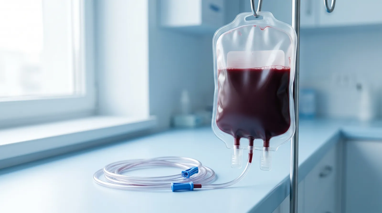
The complication, and the most important piece of information-gain on this page, is what repeated blood removal does over time. The 2024 review described phlebotomy as a "double-edged sword": each draw depletes iron, and iron depletion plus the resulting tissue hypoxia can activate the same hypoxia-inducible factor (HIF) pathway that drives EPO, potentially keeping the red-cell production signal switched on. In other words, frequent phlebotomy can become a treadmill, lowering the number on paper while the body keeps trying to rebuild, and the iron depletion itself can cause fatigue and other symptoms. The review concluded that phlebotomy should not be a reflex, that dose reduction and risk-factor management should come first, and that the decision to remove blood should be a shared one weighing the weak evidence for the thresholds against the real costs of repeated draws. This does not mean phlebotomy is never appropriate; for a genuinely high, symptomatic hematocrit it can be the right call. It means it is a considered second-line tool, not an automatic monthly ritual.
4. Monitor ferritin and iron, especially with repeated phlebotomy
Because testosterone frees up iron and phlebotomy removes it, iron status (ferritin) deserves its own line on the panel for anyone having blood drawn more than occasionally. Repeated draws can push a man into iron deficiency, which brings its own fatigue and, ironically, can perpetuate the cycle that the draws were meant to break. Some clinicians track ferritin alongside hematocrit so that they are not solving a red-cell problem by creating an iron problem. This is genuine nuance that most clinic pages skip, and it is part of why the dose-first approach is gaining ground.
5. Hydration, sleep apnea, smoking, and other contributors
Several non-testosterone factors raise hematocrit and are worth fixing in parallel, because they compound the effect and some are reversible. Dehydration concentrates the blood and can turn a borderline reading into an action-line one, so being well hydrated before a blood draw avoids a falsely high spurious result. Untreated obstructive sleep apnea is a major contributor: the repeated nighttime drops in oxygen drive EPO, and treating the apnea can meaningfully lower hematocrit. Because testosterone can itself worsen sleep apnea, this can become a self-reinforcing loop, which is why a man with high hematocrit and loud snoring or daytime sleepiness is often worth a sleep evaluation rather than another phlebotomy. Smoking raises hematocrit through carbon monoxide and chronic mild hypoxia, and stopping helps. Chronic lung disease and living or traveling at high altitude push it up too; men sensitive to this sometimes see a seasonal or travel-related bump, and a stint at altitude (a ski trip or a move to a mountain town) can show up as a higher reading at the next lab. None of these replaces dose and route management, but ignoring them makes everything else work harder, and treating a reversible contributor sometimes solves the problem without touching the testosterone at all.
Two more practical situations come up often enough to flag briefly. Surgery and significant blood loss interact with both testosterone and hematocrit, so any planned procedure is a reason to tell the surgical team you are on TRT and to coordinate timing of labs and any dose hold with your prescriber; this is a provider-coordinated decision, not something to manage alone. And the question of a chronically managed but upper-range hematocrit, sitting in the low 50s for years, is a genuine unknown: the long-term risk of a stable, well-monitored hematocrit at that level is not firmly established by outcome data, which is part of why the 2024 review urged a measured approach rather than aggressive intervention. The reasonable stance is to keep it as low as the therapy comfortably allows, monitor it, and not panic at a value that has been stable and asymptomatic for a long time.
What about supplements or medications to lower hematocrit?
There is no well-evidenced supplement that reliably lowers hematocrit, and there is no standard prescription drug used to keep men on TRT while suppressing red-cell production. You will see various supplements promoted for "thinning the blood," but the honest framing is that the evidence is weak to absent, and they are no substitute for the dose, route, and phlebotomy levers above. Off-label medication approaches exist in rare, specialist-managed situations, but they are not a first-line consumer option and are well outside the scope of self-management. The dependable tools remain flattening the peak and, when needed, clinician-ordered blood removal.
How fast does hematocrit rise, and how fast does it come back?
Hematocrit typically climbs over the first three to six months of therapy and tends to plateau within the first year, and after a phlebotomy it drops promptly but begins refilling within weeks, often returning toward its prior level over one to a few months if the underlying stimulus is unchanged. Knowing the timeline prevents both panic at an early rise and surprise when a post-phlebotomy value bounces back.
On starting or increasing testosterone, the red-cell response is not instant; it builds as the marrow ramps up, which is why guideline monitoring checks hematocrit a few months in rather than at week two. Smoother delivery (split dosing, subcutaneous, gel) tends to produce a lower plateau than a single large weekly intramuscular shot at the same total dose, which is the entire rationale for the route-and-frequency levers. After a therapeutic phlebotomy, the value falls quickly, but because the testosterone stimulus is still present, the marrow rebuilds, and the hematocrit creeps back up over the following weeks to months. This is the practical case against treating phlebotomy as a standalone solution: if nothing else changes, you are draining a tub with the tap still running. The chart below illustrates the pattern across cadence cohorts.
A decision framework: what gets done at each hematocrit level
Clinicians generally match the response to the level: keep monitoring in range, recheck and address contributors in the early-watch band, reduce dose or smooth the route as the value approaches the action line, add clinician-ordered phlebotomy for a high or symptomatic value, and refer to a hematologist when it stays high despite management or looks atypical. This is a map of the logic, not a self-treatment protocol.
The framework below summarizes how the levers above tend to be sequenced against the number. Every box is a clinician's call informed by your symptoms, risk factors, and trend, not just a single reading.
How often should hematocrit be checked?
Hematocrit is checked at baseline before starting, again at roughly three months, around six to twelve months, and periodically after that once stable, with an extra recheck after any dose, route, or schedule change. Monitoring cadence is what turns a silent rise into a caught one.
The baseline CBC matters because it tells the clinician whether you started with a high-normal hematocrit (a reason for caution) and gives every later value something to compare against. The early recheck at around three months catches the initial climb while it is still easy to manage. Once the value has plateaued and sat stable for a year or so, the interval can stretch, but it does not disappear, because contributors like new-onset sleep apnea or a dose change can move it. The single most useful habit is to recheck after any change, since that is exactly when the trend shifts. Hematocrit is only one line on a broader panel that also tracks estradiol, SHBG, PSA, and lipids; the full marker-by-marker breakdown and retest schedule live in our complete TRT bloodwork panel guide, and the estradiol side specifically in our managing estradiol on TRT guide.
When to involve a hematologist or rule out polycythemia vera
A hematologist should be involved when hematocrit stays high despite dose reduction, route changes, and contributor management, when the value is very high, or when the blood count looks atypical in a way that suggests a primary marrow disorder rather than a testosterone effect. Most TRT erythrocytosis never needs a specialist, but knowing the referral triggers matters.
The classic reason to escalate is a hematocrit that will not come down with the standard levers, or that climbs back fast and high after every intervention. Beyond persistence, certain patterns point away from a simple secondary erythrocytosis: a markedly elevated hematocrit, abnormalities in the other cell lines (high platelets or white cells, not just red cells), an enlarged spleen, or symptoms like intense itching after a hot shower. Those features raise the question of polycythemia vera, the clonal marrow disorder, and a hematologist can test for the JAK2 mutation and measure other markers to distinguish it from a testosterone-driven rise. The reason to get this right is that the two conditions are managed differently: secondary erythrocytosis is handled by adjusting the testosterone and, when needed, removing blood, while polycythemia vera is a hematologic diagnosis with its own treatment pathway. If you are managing a stubborn or unusual hematocrit, asking your clinician whether a hematology referral is warranted is a reasonable step, not an overreaction.
What real TRT trackers log
Aggregated, OCR-scanned bloodwork from our trackers shows hematocrit behaving exactly as the guidance predicts: the typical value sits in the normal range, a cohort drifts into the early-watch band, and a smaller tail crosses the action line, with smoother dosing cohorts trending slightly lower. These are the patterns behind the thresholds above.
Because the ProtocolPlus app lets users scan their lab reports, the hematocrit figures here come from real CBC values that people log rather than from a survey. In our tracking data, drawn from roughly 6,400 TRT trackers, the median hematocrit sits near 48%, comfortably in range, with the upper quartile around 52% (the early-watch zone) and about 9% crossing the ~54% action line. The injection-cadence split, once-weekly about 32%, twice-weekly or every-3.5-days about 46%, and every-other-day or daily about 22%, lines up with the mechanism: the every-other-day and split-dose cohorts trend a touch lower on hematocrit than the once-weekly cohort, consistent with the "flatter peaks, weaker red-cell stimulus" story, though this is a population tendency and not a prediction for any individual. None of these numbers is a target to chase or a medical claim; they are a snapshot of what the bloodwork-first approach is built around.
Frequently asked questions
Sources
Factual and clinical claims are sourced below. Testosterone dosing and threshold figures are described as studied in trials, defined in guidelines, or typical of clinic practice, not as recommendations.
- Endocrine Connections (2024) — Bond P, Verdegaal T, Smit DL, Testosterone therapy-induced erythrocytosis: can phlebotomy be justified? 13(10):e240283. https://pmc.ncbi.nlm.nih.gov/articles/PMC11466264/ — retrieved 2026-06-17.
- Endocrine Society (2018), JCEM — Bhasin S, et al., Testosterone Therapy in Men With Hypogonadism: An Endocrine Society Clinical Practice Guideline. 103(5):1715. https://academic.oup.com/jcem/article/103/5/1715/4939465 — retrieved 2026-06-17.
- American Urological Association (2018, amended 2023) — Mulhall JP, et al., Evaluation and Management of Testosterone Deficiency: AUA Guideline. https://www.auanet.org/guidelines-and-quality/guidelines/testosterone-deficiency-guideline — retrieved 2026-06-17.
- NEJM (2023) — Lincoff AM, et al. (TRAVERSE), Cardiovascular Safety of Testosterone-Replacement Therapy. 389:107. https://pubmed.ncbi.nlm.nih.gov/37326322/ — retrieved 2026-06-17.
- U.S. Food & Drug Administration — Testosterone products: drug safety communication and labeling on cardiovascular risk and blood pressure. https://www.fda.gov/drugs/drug-safety-and-availability/fda-drug-safety-communication-fda-cautions-about-using-testosterone-products-low-testosterone-due — retrieved 2026-06-17.
About this guide. Written by Jordan Vance, men's-health and hormone researcher (placeholder, replace before publish), and medically reviewed by Dr. Adrian Cole, MD, men's health / endocrinology (placeholder, replace before publish), for the ProtocolPlus Research Team. This guide is educational and not medical advice.